Surface Anatomy Of Ribs / Thorax Surface Anatomy 4 Edition - Superficial dissection of the back of the neck.. Gross anatomy there are 12 pairs of ribs which are separated by intercostal spaces. Some have everyday names like the palm of the hand, the sole of the foot, and the nape of the neck. Developing an understanding of the human form requires significant work and a wide range. The ribs help protect vital organs in the thorax such as the heart. The surface anatomy of the ear is frequently cut and reconstructed during mohs surgery.
Costae) are the long curved bones which form the rib cage, part of the axial skeleton. Atypical ribs rib 1 is shorter, most curved and wider than the other ribs. The heads of ribs 1 10 11 and 12 have a single facet for articulation with the bodies of the thoracic vertebrae. The intercostal muscles occupy each of the intercostal spaces and are named according to their surface relations, i.e. Learning anatomy classically involved dissection of the deceased whether directly in the laboratory or from texts, drawings, photographs or videos.
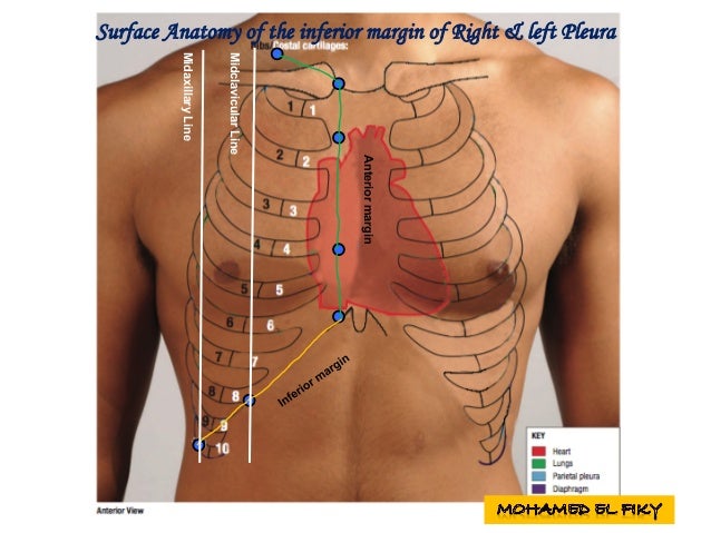
Ppt Pemeriksaan Thorax Powerpoint Presentation Free Download Id 346746 from image.slideserve.com This muscle assists in depression of the ribs. Clinical anatomy students learn to use imaginary lines and bony landmarks on the front and back of finally, three lines help describe surface locations on the back imagine drawing lines that follow the costal margins (lower borders of the anterior rib cage) and meet at the lower part of the sternum. Now notice the rib belongs to the side on which it is both ends touch the surface. Draw a line downwards and laterally from here to cut lateral border of sacrospinalis just where tip of 12th rib emerges. The ribs stretches posteriorly from thoracic vertebrae to the anterior lateral edges of the sternum. The heads of ribs 1 10 11 and 12 have a single facet for articulation with the bodies of the thoracic vertebrae. The first pair of ribs articulates with the sternum through cartilaginous joints or synchondroses and is relatively immobile. The final two pairs of ribs are floating the fibres pass superolaterally to insert into the internal surface of costal cartilages of ribs two to six.
Rib 2 is thinner and longer than rib 1, and has two articular facets on the head as normal.
This muscle assists in depression of the ribs. There are twelve pairs of ribs. Draw a line downwards and laterally from here to cut lateral border of sacrospinalis just where tip of 12th rib emerges. Rib anatomy landmarks lungs and ribs anatomy rib anatomy numbers 10th rib anatomy floating ribs anatomy thorax surface anatomy 1st rib anatomy lower rib anatomy human anatomy rib cage muscles rib cage structure typical rib anatomy single rib anatomy anterior. The surface anatomy of the ear is frequently cut and reconstructed during mohs surgery. Includes images, video, and free quiz. Gross anatomy there are 12 pairs of ribs which are separated by intercostal spaces. The ribs/costal cartilages have various attachments to the sternum. Some have everyday names like the palm of the hand, the sole of the foot, and the nape of the neck. In vertebrate anatomy, ribs (latin: Atypical ribs rib 1 is shorter, most curved and wider than the other ribs. The exceptions are the 11th and 12th ribs that don't have this surface, which enables them much higher mobility. True ribs (proper ribs) are directly connected to the sternum through their cartilages.
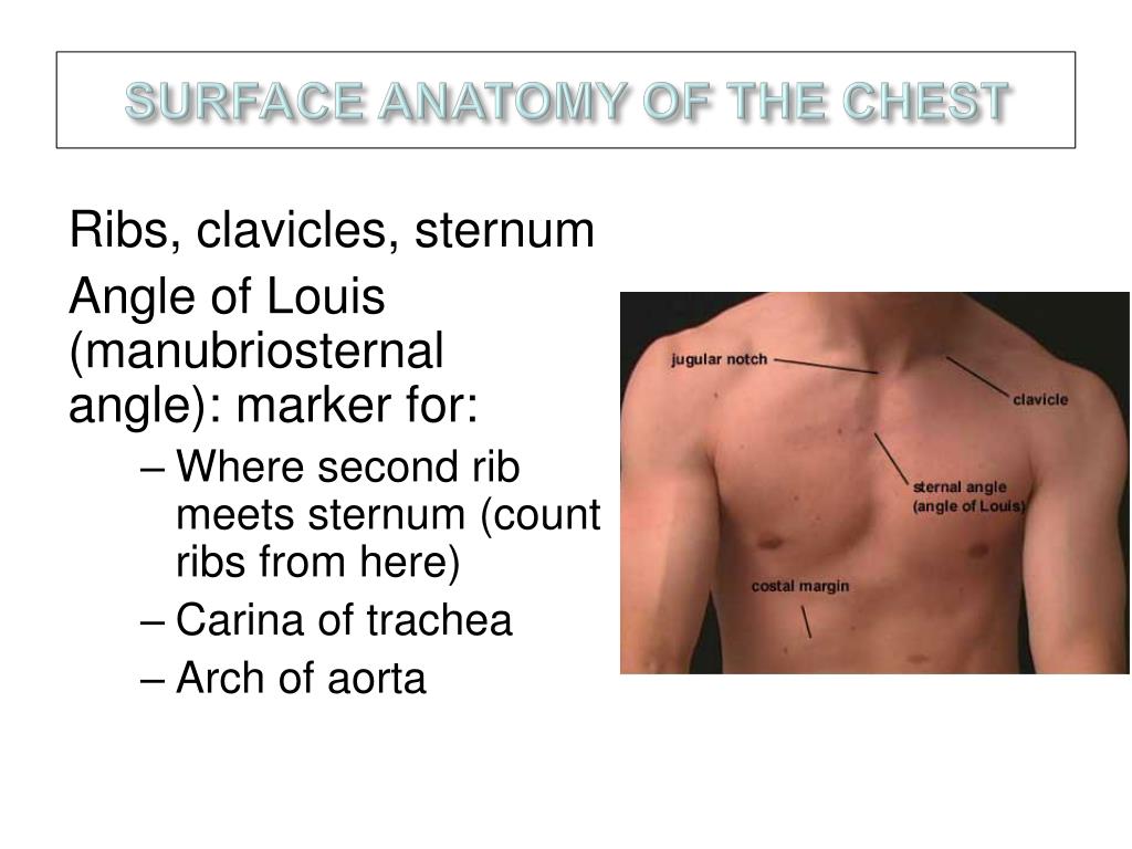
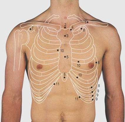
Rib anatomy landmarks lungs and ribs anatomy rib anatomy numbers 10th rib anatomy floating ribs anatomy thorax surface anatomy 1st rib anatomy lower rib anatomy human anatomy rib cage muscles rib cage structure typical rib anatomy single rib anatomy anterior. Surface markings of the thorax. Some have everyday names like the palm of the hand, the sole of the foot, and the nape of the neck. Each rib articulates posteriorly with two thoracic vertebrae by the costovertebral joint. Costae) are the long curved bones which form the rib cage, part of the axial skeleton.
Surface Anatomy Of Heart And Lungs from image.slidesharecdn.com Surface anatomy of the back. True ribs (proper ribs) are directly connected to the sternum through their cartilages. Atypical ribs rib 1 is shorter, most curved and wider than the other ribs. With the upper ribs, closer to. The recipient surface anatomy of a bony defect is typically irregular in its size and shape, which presents the clinician with a challenge as it pertains to grafting. The surface anatomy of the ear is frequently cut and reconstructed during mohs surgery. The ribs form the main structure of the thoracic cage protecting the thoracic organs, however their main function is to aid respiration3. Developing an understanding of the human form requires significant work and a wide range.
The ribs help protect vital organs in the thorax such as the heart.
The first rib surfaces looking upward and downward, and its borders inward and outward. The surface regions of the body have received their names in a variety of ways. An exception to this rule is those closest to the skin's surface run from the back of the vertebrae to the scapula eg trapezius , rhomboid s, latissimus dorsi , others wrap around the. The ribs help protect vital organs in the thorax such as the heart. The rib 1 head is small, rounded. In vertebrate anatomy, ribs (latin: Each rib articulates posteriorly with two thoracic vertebrae by the costovertebral joint. If the rib is set on the incorrect side, then only its anterior end. Learn the true ribs, false ribs, and floating ribs, as well as the difference between in this anatomy lesson, i'm going to cover the rib bones, also called costae in latin. The ribs form the main structure of the thoracic cage protecting the thoracic organs, however their main function is to aid respiration3. Superficial dissection of the back of the neck. The superior surface is marked by two grooves, which make way for the subclavian vessels. The intercostal muscles occupy each of the intercostal spaces and are named according to their surface relations, i.e.
The ribs are elastic arches of bone, which form a large part of the thoracic skeleton. .mediastinum surface anatomy thorax surface anatomy how to count ribs surface anatomy of the breast in women visualizing structures at the tiv/v the costal cartilages of ribs viii to x articulate with the inferior margins of the costal cartilages above them. Gross anatomy there are 12 pairs of ribs which are separated by intercostal spaces. The ribs stretches posteriorly from thoracic vertebrae to the anterior lateral edges of the sternum. Ribs xi and xii are called floating ribs.
Thorax Surface Anatomy 4 Edition from doctorlib.info True ribs (proper ribs) are directly connected to the sternum through their cartilages. There are twelve pairs of ribs. The rib 1 head is small, rounded. Ribs eight to ten are the false ribs and are connected to the sternum indirectly via the cartilage of the rib above them. The ribs stretches posteriorly from thoracic vertebrae to the anterior lateral edges of the sternum. Right and left scapular li. Anatomy ▶ thorax ▶ bones and cartilages ▶ the ribs. Surface anatomy and surface markings bibliographic record list of illustrations subject index.
Atypical ribs rib 1 is shorter, most curved and wider than the other ribs.
Surface anatomy and surface markings bibliographic record list of illustrations subject index. Right and left scapular li. Rib 2 is thinner and longer than rib 1, and has two articular facets on the head as normal. The recipient surface anatomy of a bony defect is typically irregular in its size and shape, which presents the clinician with a challenge as it pertains to grafting. The ribs form the main structure of the thoracic cage protecting the thoracic organs, however their main function is to aid respiration3. In vertebrate anatomy, ribs (latin: The superior surface is marked by two grooves, which make way for the subclavian vessels. Includes images, video, and free quiz. The ribs are elastic arches of bone, which form a large part of the thoracic skeleton. Anatomy ▶ thorax ▶ bones and cartilages ▶ the ribs. Atypical ribs rib 1 is shorter, most curved and wider than the other ribs. Draw a line downwards and laterally from here to cut lateral border of sacrospinalis just where tip of 12th rib emerges. In most tetrapods, ribs surround the chest, enabling the lungs to expand and thus facilitate breathing by expanding the chest cavity.
The ribs are elastic arches of bone, which form a large part of the thoracic skeleton anatomy of ribs. Superficial dissection of the back of the neck.


0 Komentar